


2026.07.20
Implant Surface Hydrophilicity Evaluation Report
Comparison of Surface Hydrophilicity Among Three Manufacturers' Implants via Water Contact Angle Measurement
Prepared by: Jeonbuk National University College of Dentistry, Dept. of Dental Biomaterials Professor Bae Tae-Seong
1. Background and Purpose of the Test
Titanium (Ti) is a highly reactive metal, so high-quality cutting fluid is used during CNC machining to prevent unwanted reactions. After machining, organic cleaning agents are used to remove the cutting fluid, but some organic residue can remain on the surface during this process. This organic residue renders the surface hydrophobic, reducing its reactivity with water and consequently delaying early bone formation (osseointegration).
Therefore, to promote osseointegration it is important to remove residual organic matter from the surface and improve its hydrophilicity. Representative methods used for this purpose include ultraviolet (UV) treatment, low-temperature plasma treatment, and femtosecond laser treatment.
This report aims to compare the surface hydrophilicity levels of implant products from three domestic and international manufacturers (Doiff, Osstem, Straumann) by measuring water contact angle, and to examine the change in contact angle before and after UV treatment. It also summarizes the principles of contact angle measurement, the osseointegration reaction mechanism of titanium implants, and the mechanism of surface hydrophilization by UV treatment, in order to aid understanding of the test results.
1.1 Principles of Water Contact Angle and Surface Hydrophilicity
Contact angle is defined as the angle formed between the surface of a water droplet placed on a solid surface and the solid surface itself, and is determined by the surface's chemical composition, surface energy, and micro-roughness. On a hydrophilic surface, the water droplet spreads widely, resulting in a small contact angle, whereas on a hydrophobic surface, the water droplet does not spread well, resulting in a relatively large contact angle.
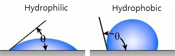
Figure 1. Conceptual diagram of water contact angle on hydrophilic and hydrophobic surfaces
1.2 Titanium Implant Surface and Osseointegration Reaction Mechanism
The surface of a titanium implant naturally forms a thin oxide layer (TiO2) in air, and this oxide layer surface contains both acidic hydroxyl groups (bridging OH) and basic hydroxyl groups (terminal OH). Because of this, the Ti surface can bind relatively easily with both acidic and basic bone-related adhesion proteins, which is known to enable faster osseointegration compared to other metals. The more hydrophilic the surface, the smoother the initial wetting and reaction with bodily fluids and proteins, allowing the reactivity of these hydroxyl groups to be fully expressed.
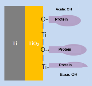
Figure 2. Schematic diagram of the binding of acidic/basic hydroxyl groups on the Ti/TiO2 surface with bone-related proteins
2. Test Materials and Methods
2.1 Test Materials
Model | Manufacturer | Surface | Lot No. |
AOBL1R501OS | Doiff, Suncheon, Korea | SLA | DF2510504 |
TS3550105 | Osstem | - | - |
021.7508 | Straumann Manufacturing, USA | SLA | ZFA75 |
2.2 Test Method
2.3 Background on the Formation of Organic Contaminants on the Implant Surface (CNC Machining Process)
In general, machined Ti implants are manufactured in the following order: CNC precision machining using cutting fluid → cleaning with an organic detergent → sterilization. During this process, the surface is finished relatively smoothly, but hydrophobic organic molecules derived from the cutting fluid and cleaning agents tend to adsorb and bond to the surface, increasing the contact angle. Therefore, a post-treatment process to decompose and remove these adsorbed organic molecules is required to improve wettability and promote osseointegration.
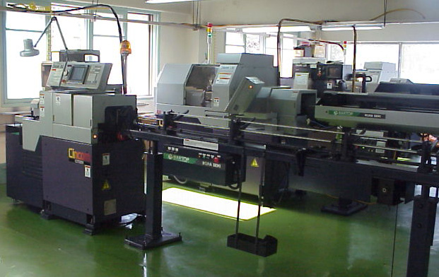
Figure 3. Ti implant CNC precision machining equipment (machining process using cutting fluid)
2.4 Mechanism of Surface Hydrophilization by UV Treatment
When the TiO2 on the SLA-treated implant surface absorbs UV light at 387 nm or shorter wavelengths, a photocatalytic reaction generates holes (h+) and free electrons (e-). The generated holes react with water molecules adsorbed on the surface to form hydroxyl groups (terminal -OH), and through strong oxidation, decompose and remove residual organic matter on the surface (C=C, C=O, C-H bonds, etc.). Meanwhile, the free electrons react with oxygen to generate reactive oxygen ions, which exhibit strong bactericidal action and also contribute to improved biocompatibility. In addition, when surface contaminants are removed and micro-roughness is formed through grit-blasting and acid-etching (SLA) treatment, the surface area and surface energy increase, and when combined with UV or low-temperature plasma treatment, a near-superhydrophilic surface can be obtained.
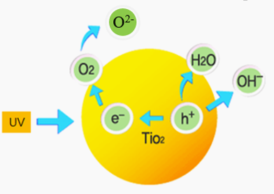
Figure 4. Mechanism of decomposition and removal of organic contaminants via the UV photocatalytic reaction on the TiO2 surface
3. Test Results
3.1 Contact Angle Comparison of SLActive Implants versus Pure Ti Plate (Reference Data)
Prior to this test, to estimate the range of contact angles according to the degree of surface treatment, the contact angle of a pure Ti plate polished with #1000 SiC abrasive paper, cleaned and dried, was compared with that of an SLActive Ti implant that had been grit-blasted and acid-etched (SLA) and specially stored. The polished Ti plate showed a contact angle of 42.3°, while the SLActive Ti implant showed 0°, indicating superhydrophilicity. This is explained by the increase in surface energy due to removal of surface contaminants and formation of micro-roughness through grit-blasting and acid-etching, and by the decomposition and removal of hydrophobic organic contaminants in the low-temperature plasma storage environment. This result serves as a reference standard for estimating the relative range (0°~100°) within which the contact angles of the three manufacturers' products fall.
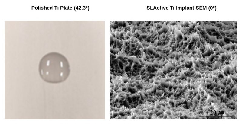
Figure 5. Comparison of the polished Ti plate (contact angle 42.3°) and SEM image of the SLActive Ti implant surface (contact angle 0°, superhydrophilic)
3.2 Results of Contact Angle Measurement Before and After UV Treatment for the Three Manufacturers' Implants
The results of water contact angle measurement before and after UV treatment for the three manufacturers' implants are as follows.
Manufacturer (Product) | Before UV Treatment (°) | After 24h UV Treatment (°) | Remarks |
Osstem | 97.29 | 97.15 | Highest contact angle among the three companies |
Doiff (OBL) | 75.68 | 63.89 | Lowest contact angle among the three companies · Best hydrophilicity |
Straumann | 82.30 | 78.69 | Intermediate level |
According to the measurement results, the Doiff (OBL) product showed 75.68° before UV treatment and 63.89° after 24 hours of UV treatment, the lowest contact angle among the three companies' products both before and after treatment. In comparison, Straumann measured 82.30°→78.69°, and Osstem measured 97.29°→97.15°, maintaining relatively higher contact angles than Doiff.
3.3 Contact Angle Measurement Photos by Manufacturer
The following are photographs of the actual contact angle measurements before and after UV treatment, taken by dropping water droplets onto the implants of the three manufacturers. The degree of droplet spreading (contact angle) can also be visually compared.
① Osstem
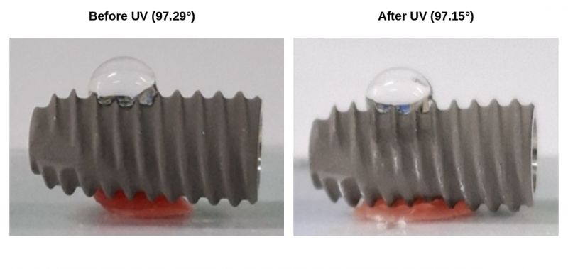
Figure 6. Comparison photo of Osstem implant contact angle before (97.29°) and after (97.15°) UV treatment
② Doiff (OBL)
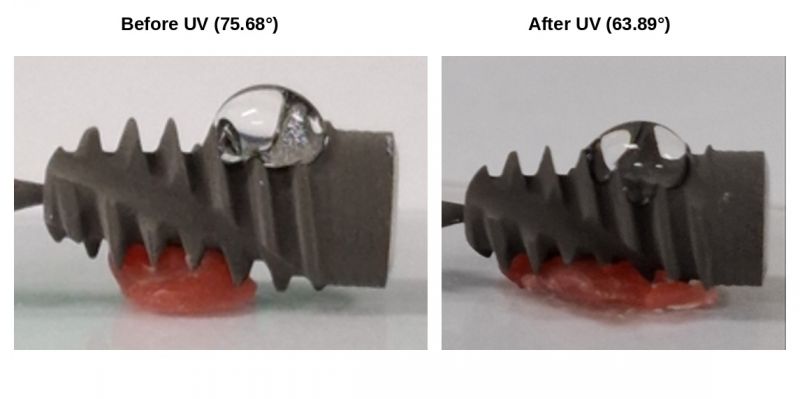
Figure 7. Comparison photo of Doiff (OBL) implant contact angle before (75.68°) and after (63.89°) UV treatment — the lowest contact angle among the three companies both before and after treatment
③ Straumann
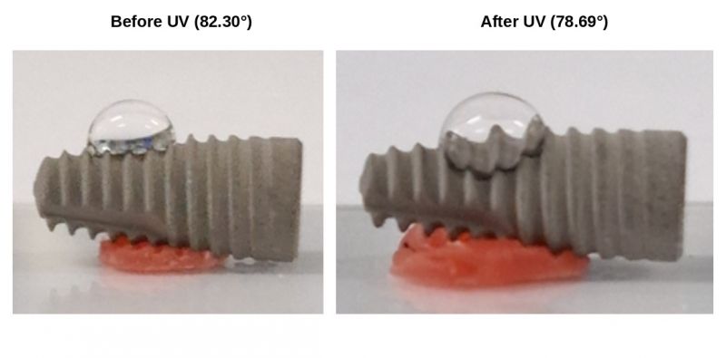
Figure 8. Comparison photo of Straumann implant contact angle before (82.30°) and after (78.69°) UV treatment
3.4 Overall Comparison of the Three Implant Types
The contact angles before and after UV treatment for the three manufacturers' products are compared side by side below. It can be visually confirmed that the water droplet on the Doiff (OBL) product spreads more widely on the surface compared to the other two companies.
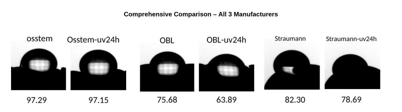
Figure 9. Overall comparison of contact angles before and after UV treatment for the three implant types (Osstem, Doiff/OBL, Straumann)
4. Discussion and Conclusion
Professor Bae Tae-Seong's Profile
Affiliation : Dept. of Dental Biomaterials, College of Dentistry, Jeonbuk National University
Position : Professor Emeritus
Field of Specialization & Research : Dental Biomaterials (Biomaterials)
Major Education & Career
Education:
D.D.S., Jeonbuk National University (1987)
M.S. in Dentistry, Jeonbuk National University (1990)
Ph.D. in Dentistry, Chosun University (2000)
Career:
Completed Residency, Dept. of Prosthodontics, Jeonbuk National University Hospital
Head, Dental Department, ROK Army Pohang Hospital
Visiting Professor, Oregon Health & Science University, USA (2001~2002)
Board Member, Korean Academy of Prosthodontics and Korean Academy of Occlusion
Director, Dental Clinical Services, Jeonbuk National University Hospital
Founding Director, Jeonbuk Center for Dental Care of the Disabled
Dean, Jeonbuk National University School of Dentistry
Major Research Achievements & Awards
Minister of SMEs and Startups Award (2017):Awarded for outstanding technology development contributing to industrial advancement through an industry-academia-research collaborative technology development project
Development of Zirconia All-Ceramic Crown Liner:Co-developed a spray-type bonding material (high-temperature liner glass) with a company (HASS Co., Ltd.) to prevent veneer coating layers from cracking or detaching in zirconia all-ceramic crowns, which are made entirely of ceramic material.
